- Home
- About Us
- Doctors
- Specialties
- Bariatric Surgery
- Bone Marrow Transplant
- Cancer
- Cardiology
- Cardiovascular And Thoracic Surgery
- Critical Care Medicine
- Dental Surgery
- Dermatology & Cosmetology
- Diabetic Foot Care
- Ear, Nose & Throat
- Endocrinology
- Fetal Medicines
- Gastroenterology
- General Medicine
- General Surgery
- HPB & Gastrointestinal Surgery
- Interventional Radiology
- IVF
- Kidney Transplant
- Laparoscopic Surgery
- Liver Transplant
- Medical And Hemato Oncology
- Neurology
- Neuro & Spine Surgery
- Nephrology And Dialysis
- Nuclear Medicine
- Orthopedic
- Ophthalmology
- Obstetrics And Gynecology
- Pathology Laboratory
- Pediatric
- Peripheral Vascular And Endovascular Surgery
- Physiotherapy and Rehabilitation
- Plastic Reconstruction
- Plastic & Cosmetic Surgery
- Pulmonary Medicine
- Radiation Oncology
- Radiology
- Robotic Surgery
- Surgical Oncology
- Urology
- Facilities
- Patient Area
- Testimonials
- Media
- Contact Us
Device Closure for ASD, VSD, and PDA
Minimally Invasive Heart Procedures
Device closure is a minimally invasive procedure used to treat specific congenital heart defects, including Atrial Septal Defect (ASD), Ventricular Septal Defect (VSD), and Patent Ductus Arteriosus (PDA). These defects involve abnormal openings in the heart’s structure, which can lead to various health issues. Device closure offers a less invasive alternative to traditional open-heart surgery, reducing recovery time and minimizing scarring.
Understanding ASD, VSD, and PDA
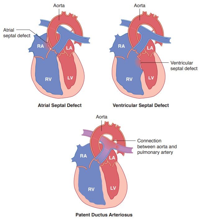
Atrial Septal Defect (ASD): ASD is a congenital heart defect where there’s an abnormal opening between the atria (the upper chambers of the heart). This allows oxygen-rich blood from the left atrium to mix with oxygen-poor blood in the right atrium, which can strain the heart and lead to complications.
Ventricular Septal Defect (VSD): VSD is a condition characterized by a hole between the ventricles (the lower chambers of the heart). This allows oxygen-rich blood to flow from the left ventricle to the right ventricle, causing the heart to work harder and potentially leading to complications.
Patent Ductus Arteriosus (PDA): PDA occurs when the ductus arteriosus, a blood vessel connecting the aorta and the pulmonary artery in a developing fetus, fails to close after birth. This can lead to abnormal blood flow and increased strain on the heart.

Device Closure Procedure

Device closure is performed under local anesthesia and involves the following steps:
1. Preparation: The patient is prepared for the procedure, and a catheter is inserted into a blood vessel, typically through the groin.
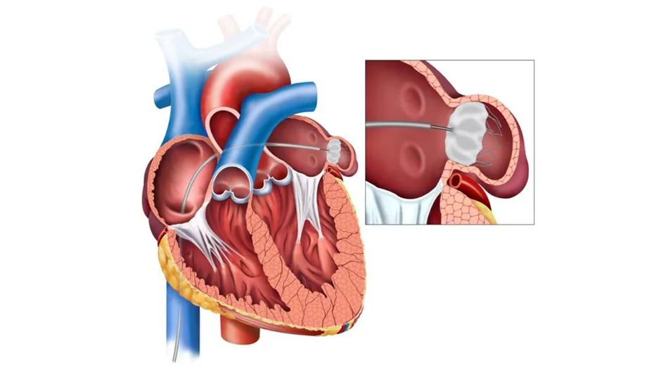
2. Guidance: Using advanced imaging techniques, such as fluoroscopy or echocardiography, the cardiologist guides the catheter to the heart defect.
3. Device Placement: A specialized device, often made of mesh or fabric, is carefully positioned within the defect.
4. Closure: The device is deployed, effectively sealing the opening. Over time, the patient’s tissues will grow around the device, permanently closing the defect.
5. Recovery: After the procedure, most patients can typically return home the same day or the day after. Recovery is relatively fast compared to traditional surgery.
Benefits of Device Closure
- Minimally Invasive: Device closure avoids the need for open-heart surgery, reducing scarring and complications associated with large incisions.
- Effective: It is an effective treatment option for many patients, improving heart function and reducing symptoms.
- Shorter Recovery: Patients often experience shorter hospital stays and quicker recovery times.

