- Home
- About Us
- Doctors
- Specialties
- Bariatric Surgery
- Bone Marrow Transplant
- Cancer
- Cardiology
- Cardiovascular And Thoracic Surgery
- Critical Care Medicine
- Dental Surgery
- Dermatology & Cosmetology
- Diabetic Foot Care
- Ear, Nose & Throat
- Endocrinology
- Fetal Medicines
- Gastroenterology
- General Medicine
- General Surgery
- HPB & Gastrointestinal Surgery
- Interventional Radiology
- IVF
- Kidney Transplant
- Laparoscopic Surgery
- Liver Transplant
- Medical And Hemato Oncology
- Neurology
- Neuro & Spine Surgery
- Nephrology And Dialysis
- Nuclear Medicine
- Orthopedic
- Ophthalmology
- Obstetrics And Gynecology
- Pathology Laboratory
- Pediatric
- Peripheral Vascular And Endovascular Surgery
- Physiotherapy and Rehabilitation
- Plastic Reconstruction
- Plastic & Cosmetic Surgery
- Pulmonary Medicine
- Radiation Oncology
- Radiology
- Robotic Surgery
- Surgical Oncology
- Urology
- Facilities
- Patient Area
- Testimonials
- Media
- Contact Us
TEE (Transesophageal Echocardiography)
A Deeper Look into Heart Health
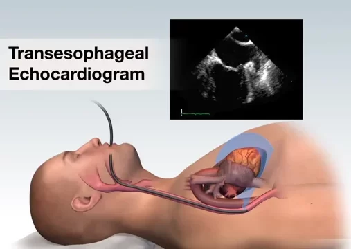
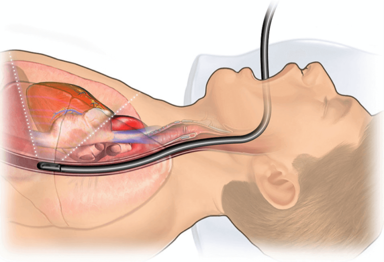
Transesophageal Echocardiography, commonly referred to as TEE, is an advanced medical imaging procedure that offers a unique and in-depth view of the heart and its function. This diagnostic tool provides cardiologists with high-resolution, detailed images of the heart by using a specialized ultrasound transducer inserted through the esophagus.
Understanding TEE
TEE involves the use of a specialized ultrasound probe, known as a transducer, which is attached to a thin, flexible tube. The procedure typically includes the following key elements:
Anesthesia: To ensure your comfort and reduce the gag reflex, a local anesthetic is applied to the throat, and you may be given a sedative to help you relax.
Transducer Insertion: The transducer is mounted on the end of the tube and carefully passed through the mouth and into the esophagus, located just behind the heart.
Ultrasound Imaging: Once in position, the transducer emits high-frequency sound waves that bounce off the heart’s structures. The echoes are converted into detailed, real-time images of the heart’s chambers, valves, and major blood vessels.
Doppler Ultrasound: TEE often includes Doppler ultrasound, which allows for the assessment of blood flow patterns, helping in the detection of conditions such as valve regurgitation or stenosis.

The TEE Procedure

Preparation: You’ll be asked to refrain from eating or drinking for a few hours before the procedure. An intravenous (IV) line may be inserted for sedation and medication.
Transducer Insertion: The TEE probe is introduced through the mouth and gently guided into the esophagus.
Imaging: High-resolution images of the heart are captured in real-time, and your healthcare provider may explain the findings as they occur.
Post-Procedure: After the TEE, you’ll be monitored as the sedation wears off, and you may need to rest for a brief period.
Why TEE is Important?
TEE is a vital component of cardiac diagnostics and care for several reasons:
Detailed Assessment: It provides exceptionally detailed images of the heart, which is particularly important for complex heart conditions and surgeries.
Diagnostic Precision: TEE aids in the precise diagnosis and assessment of heart valve problems, blood clots, congenital heart defects, and other cardiac issues.
Guiding Procedures: The real-time information obtained from TEE guides cardiologists during interventions, such as cardiac surgery or the placement of cardiac devices.
Monitoring: TEE is used to monitor the progress of treatment, ensuring that patients receive the most appropriate care.
Benefits of TEE
- High Resolution: TEE provides exceptionally clear and detailed images, aiding in precise diagnosis and treatment planning.
- In-Depth Examination: It allows for a thorough examination of cardiac structures and function.
- Guidance for Procedures: TEE provides real-time guidance for cardiac interventions, improving the accuracy of procedures.
- Minimally Invasive: While it involves a probe inserted into the esophagus, TEE is less invasive than other imaging methods.

