
Introduction
Your heart works continuously to pump blood throughout your body. But how can you know whether it is pumping properly, whether its valves are working correctly, or whether its chambers are healthy?
One of the most useful tests doctors recommend to understand the structure and function of the heart is a 2D echocardiogram, commonly known as a 2D echo. It uses ultrasound waves to create moving images of the heart, allowing doctors to examine how it beats and how blood flows through its chambers and valves.
A 2D echo test is non-invasive, does not use X-rays, and is generally painless. It can help doctors investigate symptoms such as breathlessness, chest discomfort, unusual tiredness, and swelling in the legs. It is also useful for evaluating known heart conditions and monitoring heart function over time.
Understanding what a 2D echo shows, when you may need one, and what happens during the test can help you feel more confident about your heart health.
What Is a 2D Echo Test?
A 2D echo test is a type of echocardiography that uses high-frequency sound waves to produce two-dimensional, moving images of the heart.
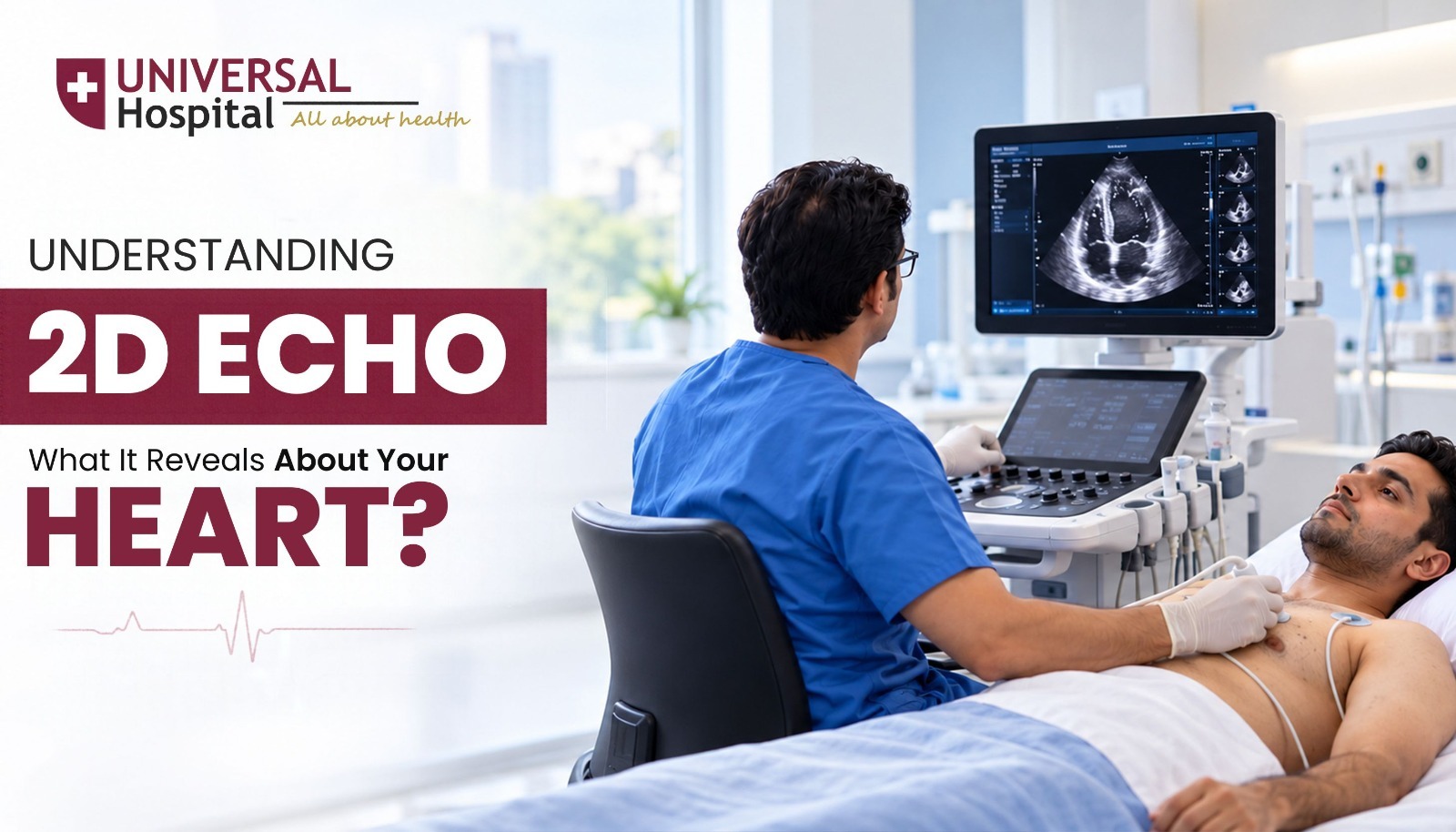
During the test, a trained healthcare professional moves a small device called a transducer over the chest. The device sends sound waves into the body and receives the echoes that bounce back from the heart’s structures. A computer converts these signals into images that the doctor can study.
These images help doctors assess:
- The size and shape of the heart.
- The thickness and movement of the heart walls.
- How effectively the heart pumps blood.
- The structure and function of the heart valves.
- Blood flow abnormalities when assessed alongside Doppler imaging.
- Signs that may suggest certain heart conditions.
Unlike an electrocardiogram (ECG), which records the heart’s electrical activity, a 2D echo provides visual information about the heart’s structure and movement.
The two tests offer different kinds of information and may be recommended together when a doctor needs a more complete assessment.
What Does a 2D Echo Reveal About Your Heart?
A 2D echocardiogram can provide valuable information about several important aspects of heart health. The findings help doctors decide whether further tests, medication, monitoring, or other treatment may be needed.
1. How Well Your Heart Pumps Blood
One of the primary purposes of a 2D echo is to evaluate the heart’s pumping function.
The left ventricle is the main chamber responsible for pumping oxygen-rich blood to the body. Doctors examine how well this chamber contracts and pushes blood into circulation.
A measurement called the ejection fraction (EF) may be calculated to estimate the percentage of blood pumped out of the left ventricle with each contraction.
For many adults, a left ventricular ejection fraction of approximately 50–70% is considered within the usual range, although interpretation depends on the measurement method, clinical context, and laboratory standards.
A reduced ejection fraction may indicate that the heart’s pumping ability is weakened. However, a normal ejection fraction does not rule out every type of heart problem, including certain forms of heart failure.
2. The Size and Structure of Heart Chambers
The heart has four chambers: two atria and two ventricles. Each chamber plays a specific role in moving blood through the heart and lungs.
A 2D echo allows doctors to examine whether these chambers are normal in size or have become enlarged.
Enlargement can occur for several reasons, including long-standing high blood pressure, valve disorders, certain heart muscle conditions, or abnormal heart rhythms.
The test can also help assess the thickness of the heart walls. For example, high blood pressure may cause the heart muscle to become thicker over time.
Identifying these structural changes can help doctors understand the possible cause of symptoms and determine whether treatment or follow-up is necessary.
3. The Condition of Your Heart Valves
Heart valves open and close to keep blood moving in the correct direction. When a valve does not function properly, the heart may have to work harder to maintain circulation.
A 2D echo can help identify structural abnormalities and assess suspected valve disease. Doppler imaging can provide additional information about blood flow across the valves.
Two common valve problems are:
- Valve stenosis: A valve becomes narrowed, restricting the flow of blood.
- Valve regurgitation: A valve does not close properly, allowing some blood to flow backwards.
Depending on the severity, valve disease may cause breathlessness, fatigue, dizziness, chest discomfort, or swelling. Some people may not experience noticeable symptoms in the early stages.
An echocardiogram helps doctors assess the type and severity of a valve problem and decide whether regular monitoring, medication, or a procedure should be considered.
4. Signs of Possible Heart Failure
Heart failure occurs when the heart cannot pump blood effectively enough or cannot fill with blood properly to meet the body’s needs.
A 2D echo is an important investigation when doctors suspect heart failure. It can assess the heart’s pumping function, chamber size, wall movement, and certain features of how the heart relaxes and fills.
Symptoms that may prompt an evaluation include:
- Breathlessness during physical activity or while lying down.
- Persistent tiredness or weakness.
- Swelling in the feet, ankles, or legs.
- Unexplained weight gain related to fluid retention.
- Reduced ability to perform everyday activities.
These symptoms can also occur because of other medical conditions. A 2D echo helps doctors investigate possible cardiac causes, but the diagnosis of heart failure usually requires a combination of symptoms, examination findings, imaging, and other tests.
5. Abnormal Heart Wall Movement
The heart muscle must contract in a coordinated way to pump blood efficiently. If a section of the heart wall moves less than expected or does not move normally, it may indicate an underlying problem.
Abnormal wall movement can sometimes be associated with damage caused by a previous heart attack or reduced blood supply to part of the heart muscle.
Doctors interpret these findings alongside symptoms, medical history, ECG results, and other investigations. A resting 2D echo alone cannot reliably rule out every form of coronary artery disease.
6. Fluid Around the Heart
A 2D echo can also detect fluid collecting in the sac surrounding the heart, a condition called pericardial effusion.
Small amounts of fluid may not cause symptoms, while larger or rapidly developing collections can interfere with the heart’s ability to fill with blood.
Depending on the amount of fluid and its effect on the heart, symptoms may include chest discomfort, breathlessness, or a rapid heartbeat.
An echocardiogram helps doctors assess the amount of fluid and whether it is affecting the heart’s function. Significant pressure on the heart requires urgent medical assessment.
When Should You Get a 2D Echo Test?
A doctor may recommend a 2D echo when symptoms, examination findings, or medical history suggest that the heart needs closer evaluation.
Common reasons include:
- Persistent or unexplained breathlessness.
- Chest discomfort when a cardiac cause is suspected.
- Swelling in the legs or ankles.
- A newly detected heart murmur.
- Suspected heart valve disease.
- Signs or symptoms of heart failure.
- Certain abnormal ECG findings.
- Monitoring a previously diagnosed heart condition.
- Assessment of some heart conditions before or after treatment.
People with high blood pressure, diabetes, or other cardiovascular risk factors may need cardiac evaluation depending on their symptoms and overall clinical condition. However, a 2D echo is not automatically necessary for every person with these risk factors.
Do not wait for a routine appointment if you experience severe or persistent chest pain, sudden breathlessness, fainting, or other symptoms that may indicate a medical emergency. Seek immediate medical care.
How Is a 2D Echo Test Performed?
If your doctor has recommended a 2D echo, there is usually no need to feel anxious. A standard transthoracic echocardiogram is a straightforward procedure that does not involve needles or surgery.
Here is what you can generally expect.
Step 1: Preparation
You may be asked to change into a hospital gown and lie on an examination bed. The healthcare professional may ask you to turn slightly onto your left side to obtain clearer images of the heart.
For a standard 2D echo, fasting is usually not required. However, follow any specific instructions provided by your hospital or doctor.
Step 2: Applying the Gel
A small amount of gel is applied to your chest. This helps the ultrasound transducer make proper contact with the skin and transmit sound waves effectively.
Step 3: Capturing Heart Images
The technician or qualified healthcare professional moves the transducer over different areas of your chest to obtain images from several angles.
You may be asked to change your position or briefly hold your breath while certain images are recorded.
The test is generally painless, although you may feel slight pressure from the device.
Step 4: Reviewing the Results
The images are reviewed and interpreted by an appropriately qualified clinician. The findings are considered alongside your symptoms, medical history, examination, and other test results.
Your doctor will explain what the report means and whether any treatment, additional investigations, or follow-up appointments are required.
A standard 2D echo often takes around 15–45 minutes, although the duration varies depending on the complexity of the examination.
Is a 2D Echo Test Safe?
Yes. A standard 2D echocardiogram is considered a safe diagnostic test for most people.
It uses ultrasound waves rather than ionising radiation, making it different from imaging tests such as X-rays and CT scans.
Key benefits include:
- Non-invasive: The standard test does not require an incision or insertion of a device into the body.
- Generally painless: Most people experience little to no discomfort.
- No radiation exposure: Ultrasound imaging does not use ionising radiation.
- Real-time imaging: Doctors can observe the heart moving and contracting.
- Useful for follow-up: It can help monitor certain diagnosed heart conditions over time.
Most people can return to their normal daily activities immediately after a standard 2D echo.
Other types of echocardiography, such as transoesophageal echocardiography, involve different preparation and may require sedation. Your doctor will explain any additional precautions if a specialised test is needed.
2D Echo vs ECG: What Is the Difference?
An ECG and a 2D echo are both valuable tools for evaluating heart health, but they examine different aspects of the heart.
| Feature | 2D Echo | ECG |
| Full form | Two-dimensional echocardiography | Electrocardiogram |
| What it examines | Heart structure, movement, and pumping function | Electrical activity and rhythm |
| Uses | Ultrasound waves | Electrodes placed on the skin |
| Helps identify | Valve abnormalities, chamber enlargement, reduced pumping function, and fluid around the heart | Abnormal rhythms and electrical changes that may suggest certain heart conditions |
| Radiation | No | No |
| Duration | Often 15–45 minutes | Usually a few minutes |
Neither test replaces the other. A doctor may recommend both to understand different aspects of a patient’s heart health.
It is also important to remember that a normal ECG or normal 2D echo does not rule out every heart condition. Further testing may be needed if symptoms persist or the clinical findings raise concern.
Understanding Your 2D Echo Report
Receiving a medical report filled with unfamiliar terms can be confusing. Knowing a few common terms can help you understand the discussion with your doctor.
Ejection Fraction (EF)
This estimates how much blood the left ventricle pumps out with each heartbeat. A reduced EF may indicate impaired pumping function, but it must be interpreted in the context of the complete examination.
Left Ventricular Function
This describes how well the main pumping chamber of the heart contracts. Reports may describe this function as normal, mildly reduced, moderately reduced, or severely reduced.
Valve Regurgitation
This means that some blood flows backwards through a valve because it does not close completely. Its significance depends on the affected valve and the severity of the leak.
Chamber Enlargement
This indicates that one or more heart chambers are larger than expected. The doctor will consider possible causes, including high blood pressure, valve disease, or abnormal heart rhythms.
Pericardial Effusion
This refers to fluid around the heart. The report may describe its size and whether there are signs that the fluid is affecting the heart’s function.
An abnormal result does not always mean that you need surgery or urgent treatment. Some findings require only periodic monitoring, while others need medication, further investigations, or specialist care. Your doctor is the right person to interpret your report and recommend the next steps.
Can a 2D Echo Detect Every Heart Problem?
Although a 2D echo provides valuable information, it cannot detect every type of heart disease.
For example, it may identify changes in heart function associated with certain cardiac conditions, but it does not directly show every narrowing or blockage in the coronary arteries.
Depending on your symptoms and medical history, your doctor may recommend additional tests, such as:
- ECG: To evaluate the heart’s electrical activity.
- Blood tests: To assess markers associated with heart muscle injury or heart failure, where appropriate.
- Stress testing: To investigate certain heart-related symptoms during physical activity or medication-induced stress.
- Coronary CT angiography or invasive coronary angiography: To examine the coronary arteries when indicated.
- Holter monitoring: To record the heart rhythm over a longer period.
The choice of test depends on the suspected condition. Your doctor will determine which investigations are appropriate rather than relying on one test alone.
How Can You Take Better Care of Your Heart?
Diagnostic tests help identify potential problems, but everyday habits also play an important role in maintaining cardiovascular health.
Consider the following measures:
- Monitor blood pressure: High blood pressure can place additional strain on the heart over time.
- Manage diabetes and cholesterol: Follow your doctor’s advice on monitoring and treatment.
- Stay physically active: Choose regular activity suited to your health and fitness level.
- Eat a balanced diet: Include vegetables, fruits, whole grains, pulses, and other nutritious foods while limiting excess salt and highly processed foods.
- Avoid tobacco: Smoking and other tobacco use increase cardiovascular risk.
- Attend recommended follow-ups: If you have an existing heart condition, follow your prescribed treatment and monitoring schedule.
- Pay attention to symptoms: Do not ignore persistent breathlessness, unusual fatigue, or unexplained swelling.
These habits support heart health, but they do not replace medical evaluation when symptoms develop.
Why Choose Universal Hospital for Heart-Related Concerns?
When you are concerned about your heart, you need clear answers, appropriate investigations, and a medical team that understands your concerns. Choosing the right hospital can help you navigate the diagnostic and treatment process with greater confidence.
At Universal Hospital, Surat, patients can seek medical evaluation for heart-related symptoms and discuss appropriate investigations with their treating doctors.
The hospital’s website provides information about its medical services and specialities. For details about echocardiography availability, appointment scheduling, and the appropriate cardiac evaluation for your condition, contact the hospital directly.
When seeking care for a possible heart condition, it is important to look for:
- Appropriate diagnostic evaluation: Tests selected according to your symptoms and medical history.
- Qualified medical guidance: Clear explanations of findings and recommended next steps.
- Coordinated care: Appropriate referrals and further investigations when required.
- Patient-centred communication: Support in understanding reports, treatment options, and follow-up requirements.
- Timely medical attention: Prompt assessment when symptoms suggest an urgent problem.
Whether you have been advised to undergo a 2D echo or are concerned about unexplained symptoms, discussing your concerns with a qualified doctor is an important first step.
Conclusion
A 2D echo is an important diagnostic tool that helps doctors understand how the heart is structured and how effectively it works. From evaluating pumping function and heart valves to identifying chamber enlargement and fluid around the heart, the test provides valuable information for diagnosing and monitoring several cardiac conditions.
However, the results should always be interpreted alongside your symptoms, medical history, and other relevant investigations. Even when an earlier test is normal, new or persistent symptoms deserve appropriate medical attention.
Taking care of your heart begins with recognising warning signs, attending recommended check-ups, and seeking timely medical advice.
Take the Next Step Towards Better Heart Health
If you have been advised to undergo a 2D echo or are experiencing symptoms such as breathlessness, unusual fatigue, or suspected heart-related discomfort, do not delay seeking medical advice.
Contact Universal Hospital, Surat, to enquire about cardiac evaluation, 2D echo testing, and the appropriate next steps for your health.
Your heart deserves careful attention, accurate diagnosis, and compassionate care. Take the first step towards understanding your heart health today.

Comment 1